- Issues
- About the Journal
- News
- Cooperation
- Contact Info
Issue. Articles
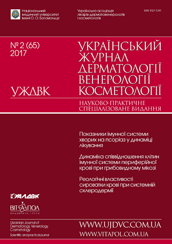
¹2(65) // 2017

1. Scientific researches
|
Notice: Undefined index: pict in /home/vitapol/ujdvc.vitapol.com.ua/en/svizhij_nomer.php on line 76 State of immune system of the body in patients with psoriasis in the dynamics of treatmentR.L. StepanenkoO.O. Bogomolets National Medical University, Kyiv |
|---|
Materials and methods. Under our complex examination were 207 patients with vulgar psoriasis, including 126 (61 %) men and 81 (39 %) women. The age of the examined patients ranged from 18 to 79 years. The indices of cellular immunity in the body were assessed by the changes in the relative and absolute number of T-(CD3+) and B-(CD19+)-lymphocytes, as well as subpopulations of (CD4+, CD8+, CD16+, CD25+CD71+, CD30+, CD95+). To identify the surface structures of lymphocytes, a direct immunofluorescence method was used in which a fluorescent label was attached to anti-CD mononuclear antibodies of the Leu series of Becton Dikinson company (USA). The calculations were performed on a laser flow cytofluorimeter of Baston company (USA).
Results and discussion. The most positive effect on the cellular part of the body’s immune system was found in patients with psoriasis who received immunotherapy with åtanercept in combination with narrow-band (311 nm) phototherapy. After treatment of patients of this group, due to a significant reduction in production of TNF and its concentration in the blood serum, there was decrease in proinflammatory changes in the cellular part of the immune system, and the number of lymphocytes with an early activation marker which initiated a further cytokine cascade of development and prolongation of the filling. In addition, a significant decrease in the number of CD30+-lymphocytes is found to be an indirect factor which indicates the switching of Tx2 responses (autoimmune manifestations) to Tx1, and therefore achievement of clinical and immunological remission of the psoriatic process. It is also very important for the functioning of the immune system to consider the decrease in the content of activated lymphocytes expressing the Fas-receptor, because due to increased apoptosis of lymphocytes, namely T-cytotoxic lymphocytes, autoimmune and proliferative changes occured in the exacerbation of the psoriatic process.
Conclusions. It has been established that all our psoriasis therapy regimens have immunorehabilitation properties, but the degree of their expressiveness was different. The most effective therapy scheme is the combined use of åtanercept and UVB which reduced proliferative and inflammatory changes in the skin, antigen load, level of CIC, autosensitivity and autoimmune disorders.
Keywords: psoriasis, systemic immunosuppressive therapy, immunological changes.
![]() To download
To download
full version need login
Original language: Ukrainian
2. Scientific researches
|
Notice: Undefined index: pict in /home/vitapol/ujdvc.vitapol.com.ua/en/svizhij_nomer.php on line 76 Dermatological and neurological features of the development of the first and second type recurrent herpes infectionU.V. Fedorova, O.O. SyzonDanylo Halytsky Lviv National Medical University, Lviv, Ukraine |
|---|
Materials and methods. We examined 120 RHI patients with 1 and 2 types of herpes simplex virus (HSV) including 41 men and 79 women. 62 patients were under age 35, 37 patients were 35—50, 21 patients were over 50.
Results and discussion. Credible gender dependence on the nature of common infection manifestations was not found except for fatigue, which was observed more frequently in women (71.0 ± 13.5 %; ð < 0.05). Gender differences were observed in the range of dermatological manifestations such as lips swelling (14.9 ± 2.97 %; ð < 0.05). Other body parts swelling (20.2 ± 4.27 %; ð < 0.01) was more significantly expressed in women however rashes on genitals (23.9 ± 3.94 %, ð < 0.05) were more common in men. Among the neurological manifestations, polyneuropathy (11.0 ± 3.03 %; ð < 0.05), sciatic neuritis (10.3 ± 3.35 %; ð < 0.05) and especially headaches (21.4 ± 5.68 %; ð < 0.05) were evidentially more frequent in women than in men. Fatigue was mostly present in patients (81.8 ± 17.2 %; ð < 0.05) who had 1 and 2 types of DNA HSV in saliva. (39.4 ± 5.83) % of them were under age 35 (ð < 0.05) and (29.6 ± 6.82) % were 35 to 50 years old (ð < 0.05). Joint pains were more manifested in patients aged 35—50 years (2.75 ± 1.19) % (ð < 0.05). Body rashes prevailed among other dermatological manifestations (24.2 ± 5.43) % in patients under age 35 (ð < 0.05). Genital rashes were detected in (18.9 ± 6.4) % of patient aged 35—50 years (ð < 0.05). Among DNA virus positive individuals the most common were dermatological manifestations such as swelling of lips (16.8 ± 4.94 %; ð < 0.01) and other body parts (20.5 ± 3.88 %; ð < 0.05), genital rashes (24.5 ± 8.03 %; ð < 0.05). Neurological manifestations were more common in patients with 1 and 2 types of DNA HSV and expressed as polyneuropathy (12.1 ± 3.15 %; ð < 0.05), eye redness (9.6 ± 4.89 %; ð < 0.05) and sciatic neuritis (11.3 ± 1.22 %; ð < 0.01).
Conclusions. Clinical manifestations of recurrent HSV infection of 1 and 2 types were mostly characterized with chronic fatigue syndrome and oedematic, dermatological and neurological disorders. The most significant clinical variations were observed in patients with active replicative ability of HSV of 1 and 2 types in saliva.
Keywords: recurrent herpes infection, herpes simplex virus of 1 and 2 types, clinical picture.
![]() To download
To download
full version need login
Original language: Ukrainian
3. Scientific researches
|
Notice: Undefined index: pict in /home/vitapol/ujdvc.vitapol.com.ua/en/svizhij_nomer.php on line 76 Dynamics of ratios of peripheral blood cells of the immune system in patients with mycosis fungoides during combined therapyL.M. HamadehÎ.Î. Bogomolets National Medical University, Kyiv |
|---|
Materials and methods. Under our supervision, there were 12 patients with mycosis fungoides (MF) who underwent treatment at the clinic of the Department of Dermatology and Venereology of O.O. Bogomolets National Medical University. The diagnosis and stages of the disease were confirmed on the basis of clinical symptoms and peripheral blood analysis. Clinical information was obtained during the analysis of patients’ medical records. Patients were treated by preparations based on prospidium chloride (chloroxiperazine) in the form of powder for injection (100 mg in 1 ampoule) and 30% ointment. Flow cytometry was performed on blood samples taken from 12 patients with MF aged 65 to 85 years in the period from 2013 to 2017. 6 patients had stage IA of MF and 6 – stage IIB. The blood test was performed 2 times before and after chemotherapy for prospidium chloride. Statistical analysis was performed using version 1.0 of SigmaSat software (Jandel Corporation, San Rafael, CA). The proportions were compared with the use of MannWhitney U test and the Spearman correlation coefficient. For all statistical tests, statistical significance was confirmed at p less than 0.05.
Results and discussion. In the blood of patients with MF of IA and IB stages, a significant decrease in the number of CD45+CD5+CD19– and CD45+CD3+ lymphocytes was detected with flow cytometry after treatment with prospidium chloride. A more pronounced decrease in the amount of CD45+CD3+CD4+CD8– T-lymphocytes of helpers was revealed in the group of patients at stage IA.
Conclusions. The study of the dynamics of changes in the quantitative composition of the helper subpopulation during therapy with the use of the prospidium chloride preparation indicated a decrease in CD45+CD4+CD45RA+CD45RO– naive Thelpers and CD45+CD4+ CD45RA–CD45RO+ Ò-cells of memory in all groups of patients. The most pronounced decrease in CD45+CD4+ CD45RA+CD45RO– naive Thelpers and CD45+CD4+CD45RA–CD45RO+ Tcells of memory was observed in patients with MF at stage IA. In patients with MF at stage IIB, in contrast to patients at stage IA, there was a significant decrease in populations of CD45+C19+CD5+CD27+ Bmemory cells and CD45+CD3–CD16+CD56+ natural killer cells.
Keywords: mycosis fungoides, combined therapy, changes in immunophenotypes of peripheral blood cells.
![]() To download
To download
full version need login
Original language: Russian
4. Scientific researches
|
Notice: Undefined index: pict in /home/vitapol/ujdvc.vitapol.com.ua/en/svizhij_nomer.php on line 76 Rheological properties of blood serum at limited and diffuse forms of systemic sclerosisO.V. Syniachenko1, V.Ya. M³kuksts1, M.V. Yermolaeva1, Ye.D. Iegud³na21 Donetsk National Medical University, Lyman |
|---|
Materials and methods. The study included 63 patients with SS aged from 16 to 67 years (mean 42 years), among them 11 % men and 89 % women. 43 % of patients had a limited form of the disease, 57 % — a diffuse form. The duration of the disease was 11 years. The I level of activity of the pathological process was registered at 41 % of patients, II — at 38 %, III — at 21 %, antitopoisomerase 1 antibodies were detected in serum at 78 % of cases, antibodies to native deoxyribonucleic acid — at 64 %, to cardiolipin — at 18 %. RPB was assessed by interfacial tensiometry with the determination of surface parameters. For control the parameters were studied in 52 healthy people and 42 patients with limited scleroderma. Skin microincisional biopsy was performed in 37 patients.
Results and discussion. SS is accompanied by violations of RPB, which are manifested by increased viscoelasticity module parameters in 8 % of patients and the surface tension in – in 66 % against the background of reducing surface elasticity level in 37 %. The last two parameters of interfacial tensiometry differ not only from those in healthy people, but also in patients with limited scleroderma. Changes in the viscoelastic properties of blood are more prevalent in diffuse form of the disease than in the limited form when there is dispersive correlation between RPB and the severity of skin lesions and extradermal manifestations of SS (heart, lung, kidney disease). The elastic and relaxation serum characteristics have predictive value in case of limited and diffuse forms of the disease.
Conclusions. SS occurs with disorders of serum level of RPB which are more characteristic to the diffuse form of the disease and are closely related to the severity of skin and internal organs lesions, the indicators of which can be used to improve the assessment of the clinical course severity and prognosis of the individual signs of the disease.
Keywords: systemic scleroderma, clinical course, blood, serum, rheology.
![]() To download
To download
full version need login
Original language: Russian
5. Scientific researches
|
Notice: Undefined index: pict in /home/vitapol/ujdvc.vitapol.com.ua/en/svizhij_nomer.php on line 76 Algorithm of acne treatment with consideration of pathogenetic componentsA.V. PetrenkoP.L. Shupyk National Medical Academy of Postgraduate Education, Ministry of Healthcare of Ukraine, Kyiv |
|---|
Meterials and methods. Results of the study are based on data of molecular genetic examination and treatment of 78 patients with acne of medium and severe degrees. Patients were divided into two groups according to treatment prescribed to them. Each group was also divided into two subgroups depending on the severity of the disease.
Results and discussion. In acne patients receiving systemic therapy with nicotinamide riboside there were significantly better outcomes compared to those receiving only local therapy. The best therapeutic effect of nicotinamide riboside was observed in patients with mutations in one or more genes.
Conclusions. Therapy with nicotinamide riboside had positive effect on course of disease in patients with gene mutations.
Keywords: acne, nicotinamide riboside, TLR4, IL 1β, IL 8.
![]() To download
To download
full version need login
Original language: Ukrainian
6. TO HELP PRACTICING PHYSICIANS
|
Notice: Undefined index: pict in /home/vitapol/ujdvc.vitapol.com.ua/en/svizhij_nomer.php on line 76 Prospects for inclusion of natural herbal remedies in the diet of patients with acne to improve skin conditionB.G. Kogan, Î.S. Svyryd-DzyadykevychÎ.Î. Bogomolets National Medical University, Kyiv |
|---|
Keywords: acne, etiology, treatment, Propionibacterium acnes, antiinflammatory effect.
![]() To download
To download
full version need login
Original language: Ukrainian
7. TO HELP PRACTICING PHYSICIANS
|
Notice: Undefined index: pict in /home/vitapol/ujdvc.vitapol.com.ua/en/svizhij_nomer.php on line 76 Complex differentiated approach to treatment of psoriasis with consideration of concomitant pathology of digestive systemT.O. Lytynska, V.I. StepanenkoÎ.Î. Bogomolets National Medical University, Kyiv |
|---|
Materials and methods. 272 patients with psoriasis aged 18 to 72 years were under observation. With consideration of the concomitant gastroenterological pathology diagnosed in patients with psoriasis and in order to evaluate the effectiveness of therapy, patients were divided into equivalent clinical groups. Patients received complex differentiated therapy taking into account the concomitant pathology of the digestive organs, namely: eradication of Helicobacter pylori infection (H. pylori infection), correction of functional disorders of the hepatic system and intestinal microbiota. Patients of the comparison group were prescribed basic therapy.
Results and discussion. Eradication of H. pylori infection and normalization of the functional state of the liver and intestine can improve the effectiveness of basic therapy, increase the duration of remission and reduce the frequency of recurrence of dermatosis in patients with psoriasis and concomitant pathology of the digestive system.
Conclusions. Complex individualized therapy, with consideration of the concomitant pathology of the digestive organs, including eradication of H. pylori infection, correction of functional disorders of the hepatic system and intestinal microbiota, allows increasing the effectiveness of the basic treatment and duration of remission as also reducing the frequency of relapses of dermatosis.
Keywords: psoriasis, Helicobacter pylori infection, hepatic system, intestinal microbiota, dysbiosis.
![]() To download
To download
full version need login
Original language: Ukrainian
8. TO HELP PRACTICING PHYSICIANS
|
Notice: Undefined index: pict in /home/vitapol/ujdvc.vitapol.com.ua/en/svizhij_nomer.php on line 76 The use of composition of «Teobon-dithiomycocide» with gentamicin for topical treatment of dermatoses complicated by fungal or bacterial microfloraV.I. Stepanenko1, L.M. Shkaraputa2, L.O. Naumova1, L.O. Tyshchenko2, L.A. Shevchenko2, Ya.V. Tsekhmister1, V.P. Kukhar2, V.A. Golikov31 O.O. Bogomolets National Medical University, Kyiv |
|---|
Materials and methods. We observed 43 patients with dermatoses complicated by fungal or bacterial infection, aged 18 to 55 years. 13 patients were diagnosed with infectious eczema, 23 — with dermatomycosis of the feet (intertrigo or dyshidrotic form), 7 – with rosacea (papulopustular form).
Results and discussion. The composition of «Teobon-dithiomycocide» with gentamicin is effective for topical treatment of certain dermatoses complicated with bacterial infection, particularly, infectious eczema, dermatomycosis of the feet (dyshidrotic form), rosacea. Therapeutic efficacy, absence of side effects and good tolerability of this drug were proved, which indicates the expediency of its use as an alternative for topical treatment of dermatoses complicated with bacterial or fungal infection.
Conclusions. The composition of «Teobon-dithiomycocide» with gentamicin is an effective alternative for topical treatment of dermatoses complicated with bacterial or fungal infections.
Keywords: «Teobon-dithiomycocide», gentamicin, topical drug, complicated dermatoses.
![]() To download
To download
full version need login
Original language: Ukrainian
9. PHARMACOTHERAPY IN DERMATOLOGY AND VENEREOLOGY
|
Notice: Undefined index: pict in /home/vitapol/ujdvc.vitapol.com.ua/en/svizhij_nomer.php on line 76 Experience of therapy of patients with resistant and heavy forms of acne and rosacea with the use of the systemic LIDOSE isotretinoinL.Ya. FedorychUkrainian Military Medical Academy, Kyiv |
|---|
Materials and methods. In the period of 2015—2017 we observed 35 patients with resistant and severe clinical forms of acne and rosacea treated with LIDOSE isotretinoin.
Results and discussion. Positive effect of LIDOSE isotretinoin therapy was registered in all 35 patients. To treat patients with severe clinical forms of acne and rosacea, a special treatment regimen using increased doses of LIDOSE isotretinoin has been proposed and applied, according to which the drug is prescribed for a short period of time at a dose of 1 mg per kg of body weight per day, with a gradual decrease in the daily dose to a minimum value. At the same time, it is mandatory to achieve the maximum LIDOSE isotretinoin dose of 120 mg per kg of body weight for the entire period of treatment, which usually takes 5—6 months.
Conclusions. Based on our own experience, systemic LIDOSE isotretinoin technology is a highly effective treatment for severe clinical forms of rosacea, acne of medium, severe and very severe degrees as also treatmentresistant forms of the disease. For treatment of these patients we suggested and applied a special treatment regimen using high doses of LIDOSE isotretinoin.
Keywords: acne, rosacea, treatment, LIDOSE isotretinoin.
![]() To download
To download
full version need login
Original language: Ukrainian
10. PHARMACOTHERAPY IN DERMATOLOGY AND VENEREOLOGY
|
Notice: Undefined index: pict in /home/vitapol/ujdvc.vitapol.com.ua/en/svizhij_nomer.php on line 76 Experience of treatment of mycotic infections of the scalpZh.V. Korolova, V.M. BorovykovP.L. Shupyk National Medical Academy of Postgraduate Education, Ministry of Healthcare of Ukraine, Kyiv |
|---|
Keywords: mycotic infections of the scalp, adolescents, systemic treatment, terbinafine.
![]() To download
To download
full version need login
Original language: Ukrainian
11. PHARMACOTHERAPY IN DERMATOLOGY AND VENEREOLOGY
|
Notice: Undefined index: pict in /home/vitapol/ujdvc.vitapol.com.ua/en/svizhij_nomer.php on line 76 Plazmotherapy (P-PRP-therapy): modern approach to treatment of atrophic scars postacneYa.O. Sulik, Î.S. Svyryd-DzyadykevychO.O. Bogomolets National Medical University, Kyiv |
|---|
Materials and methods. The review and analysis of domestic and foreign publications on the method of P-PRP-therapy for scars postacne. Our study included 11 patients (aged from 18 to 34 years) with atrophic scars postacne (8 women and 3 men). The course of P-PRP-therapy was carried out on an empty stomach with a diet for 3 days once every 2 weeks for 2 months. 9—10 ml of plasma enriched with platelets were injected subcutaneously.
Results and discussion. Using P-PRP-therapy in the treatment of atrophic scars postacne of third and fourth degree allowed achieving significant aesthetic effect manifested as the decrease of relief of the skin in the areas of lesion on the face and softening of the scars. The color and texture of the skin markedly improved at the end of the first month of treatment. After the treatment, cosmetic defects on the skin of the face were easily masked with the help of cosmetics.
Conclusions. Sufficiently high therapeutic and cosmetic efficacy and good tolerability of plasma therapy (P-PRP-therapy) in the treatment of atrophic scars postacne were established.
Keywords: plazmotherapy (P-PRP-therapy), acne, atrophic scars, postacne.
![]() To download
To download
full version need login
Original language: Ukrainian
12. Reviews
|
Notice: Undefined index: pict in /home/vitapol/ujdvc.vitapol.com.ua/en/svizhij_nomer.php on line 76 Chemical peeling in dermatology. Part II. Practical application, complications and their managementC. DiehlUniversitá Degli Studi Guglielmo Marconi, Rome, Italy |
|---|
Keywords: Chemical peeling, glycolic acid, trichloracetic acid, salicylic acid, phenol peeling, pre-peeling, post-peeling, side effects, complications.
Additional:
Dr. Christian Diehl, Department of Dermatology, Universitá Degli Studi Guglielmo Marconi
Via Plinio, 44, 00193, Rome, Italy. Å-mail: chdiehl@hotmail.com
Notice: Undefined index: attach in /home/vitapol/ujdvc.vitapol.com.ua/en/svizhij_nomer.php on line 177
Original language: English
13. Reviews
|
Notice: Undefined index: pict in /home/vitapol/ujdvc.vitapol.com.ua/en/svizhij_nomer.php on line 76 Skin manifestation of diabetesL.Î. Naumova, L.Î. Prystupiuk, V.M. KonakhO.O. Bogomolets National Medical University, Kyiv |
|---|
Materials and methods. Retrospective analysis was conducted of avaliable literature on the problem of skin lesions in patients with diabetes.
Results and discussion. Diabetes is a chronic metabolic disease. According to the International Diabetes Federation, about 285 mln. adults worldwide suffer from diabetes. It can lead to disruption of homeostasis in the skin, which is manifested by the development of various dermatoses. At least onethird of all patients with diabetes mellitus have abnormal skin changes.
Conclusions. In the vast majority of patients with diabetes, pathological changes occur on the skin. In some cases, skin manifestations may precede the diagnosis of diabetes, and in some cases they are a marker of the state of compensation for diabetes. Ñooperation of dermatologists and endocrinologists is mportant for preventing the development of severe skin lesions in diabetic patients.
Keywords: diabetes, skin lesions.
![]() To download
To download
full version need login
Original language: Ukrainian
14. Reviews
|
Notice: Undefined index: pict in /home/vitapol/ujdvc.vitapol.com.ua/en/svizhij_nomer.php on line 76 Estimation of endogenous nitric oxide metabolites in development of pathological conditions of organism. Study of nitric oxide metabolites level in blood and skin microcirculation of patients with true eczemaV.V. HiliukO.O. Bogomolets National Medical University, Kyiv, Ukraine |
|---|
Materials and methods. 48 patients (25 males and 23 females) with true eczema aged 18 to 67 were under study. In order to determine the nitric oxide metabolites level in blood plasma we used a spectrophotometric method, based on the reaction of nitrite with Griess reagent. The state of skin microcirculation was assessed using laser Doppler flowmetry.
Results and discussion. We observed the increased content of the final stable metabolites of nitric oxide in the peripheral blood of patients with true eczema. The most significantly reliable growth of indicators in comparison with healthy people was found in patients with dyshidrotic eczema that persisted for more than 10 years. We registered reduction of skin microcirculation level in patients with acute and subacute stages of eczema, and growth of this parameter in patients with chronic eczema, when compared to healthy people. The most significantly reliable skin circulation disorder was observed in patients with eczema that persisted for more than 10 years. There is a proved correlation between the level of the nitric oxide metabolites levels and the skin microcirculation disorder in affected patients.
Conclusions. The impact of the level of endogenous nitric oxide production in the blood of patients with true eczema and microcirculation disorders in the skin on the nature and severity of the clinical course of this dermatosis has been established, which must be taken into account when developing complex therapy.
Keywords: metabolites of endogenous nitric oxide, blood, skin microcirculation, true eczema.
![]() To download
To download
full version need login
Original language: Ukrainian
15. Reviews
|
Notice: Undefined index: pict in /home/vitapol/ujdvc.vitapol.com.ua/en/svizhij_nomer.php on line 76 Review of current theories of etiology and pathogenesis of facial skin hypermelanoses treatment methodsYe.I. Shelemba 1, 2, V.O. Tsepkolenko1, 31 P. L. Shupyk National Medical Academy of Postgraduate Education, Ministry of Health of Ukraine, Kyiv |
|---|
Keywords: hypermelanoses, melasma, pathogenesis, the effect of ultraviolet radiation, dermal factors microNA.
List of references:
1. Abdel-Malek Z., Suzuki I., Tada A. et al. The melanocortin‑1 receptor and human pigmentation // Ann. N. Y. Acad. Sci. — 1999. — Vol. 885. — P. 117—133.
2. Abdel-Malek Z., Swope V., Smalara D. et al. Analysis of the UV-induced melanogenesis and growth arrest in human melanocytes // Pig. Cell Res. — 1994. — Vol. 7. — P. 32.
3. Achar A., Rathi S. Melasma: a clinic-apidemiolodical study of 312 cases // Indian J. Dermatol. — 2011. — Vol. 56. — P. 380—382.
4. Bak H., Lee H., Chang S. Increased expression of nerve growth factor receptor and neural endopeptidase in the lesional skin of melisma // Dermatol. Surg. — 2009. — Vol. 35. — P. 1244—1250.
5. Budamakuntla L., Loganathan E., Suresh D. et al. A randomized, open-label, comparative study of tranexamic acid microinjections and tranexamic acid with micro-needling in patients with melisma // J. Cutan. Aesthet. Surg. — 2013. — Vol. 6. — P. 139—143.
6. Cardinali G., Kovacs D., Picardo M. Mechanisms underlying post-inflammatory hyperpigmentation: lessons from solar lentigo // Ann. Dermatol. Venereol. — 2012. — Vol. 139. — P. 148—152.
7. Chen M., Wang W., Jia W. et al. Three-dimensional contrast-enhanced sonography in the assessment of breast tumor angiogenesis: correlation with microvessel density and vascular endothelial growth factor expression // J. Ultrasound Med. — 2014 — Vol. 33. — P. 835—846.
8. Dong C., Wang H., Xue L. et al. Coat color determination by miR‑137 mediated down-regulation of microphthalmia-associated transcription factor in a mouse model // RNA-2012. — Vol. 18. — P. 1679—1686.
9. Dynoodt P., Mestdagh P., Van Peer G. et al. Identification of miR‑145 as a key regulator of the pigmentary process // J. Invest. Dermatol. — 2013 — Vol. 133. — P. 201—209.
10. Eimpunth S., Wanitphadeedecha R., Triwongwaranat D. et al. Therapeutic outcome of melasma treatment by dual-wavelength (511 and 578 nm) laser in patients with skin phototypes III—V // Clin. Exp. Dermatol. — 2014. — Vol. 39. — P. 292—297.
11. Friedmann P., Gilchrest B. Ultraviolet radiation directly induces pigment proliferation by cultured human melanocytes // J. Cell Physiol. — 1987. — Vol. 133. — P. 88—94.
12. Foldes E. Pharmaceutical effect of contraceptive pills on the skin // Int. J. Clin. Pharmacol. Ther. Toxicol. — 1988. — Vol. 26. — P. 356—359.
13. Ghosh M., Thompson D., Weigel R. PDZK1 and GREB 1 are estrogen-regulated genes expressed in hormone-responsive breast cancer // Cancer Res. — 2000 — Vol. 60. — P. 6367—6375.
14. Gilchrest B., Park H., Eller M. Mechanisms of ultraviolet light-induced pigmentation // Photochem. Photobiol. — 1996. — Vol. 63. — P. 1—10.
15. Goh C., Dlova C. A retrospective study on the clinical presentation and treatment outcome of melasma in a tertiary dermatological referral centre in Singapore// Singapore Med. J. — 1999. — Vol. 40. — P. 455—458.
16. Guerrero D. Dermocosmetic management of hyperpigmentations // Ann. Dermatol. Venereol. — 2012. — Vol. 139. — P. 166—169.
17. Guinot C., Cheffai S., Letreille J. et al. Aggravating factors for melasma: a prospective study in 197 Tunisian patients. — 2010. — Vol. 24. — P. 1060—1069.
18. Ha T. The role of microRNAs in regulatory T cells and in the immune response // Immune Netw. — 2011. — Vol. 11. — P. 11—41.
19. Hachiya A., Kobayashi A., Yoshida Y. et al. Biphasic expression of two paracrine melanogenic cytokines, stem cell factor and endothelin‑1, in ultraviolet B-induced human melanogenesis // Am. J. Pathol. — 2004. — Vol. 165. — P. 2099—2109.
20. Hamilton J. Significance of sex hormones in tanning of the skin in women // Proc. Soc. Exp. Biol. Med. — 1941. — Vol. 40. — P. 502—503.
21. Handel A., Lima P., Tonolli V. et al. Risk factors for facial melasma in women: a case-control study // Br. J. Dermatol. — 2014. — Vol. 171. — P. 588—594.
22. Handel A., Miot L., Miot H. Melasma: a clinical and epidemiological review // An. Bras. Dermatol. — 2014. — Vol. 89. — P. 771—782.
23. Hernandez-Barrera R., Torrez-Alvarez B., Castanedo-Cazares J. et al. Solar elastosis and presence of mast cells as key features in the pathogenesis of melisma // Clin. Exp. Dermatol. — 2008. — Vol. 33. — P. 305—308.
24. Hexsel D., Lacerda D., Cavalcante A. et al. Epidemiology of melasma in Brazilian patients: a multicenter study // Int. J. Dermatol. — 2014. — Vol. 53. — P. 440—444.
25. Ho S., Chan H. The Asian dermatologic patient: review of common pigmentary disorders and cutaneous diseases // Am. J. Clin. Dermatol. — 2009. — Vol. 10. — P. 153—168.
26. Imokawa G., Miyagishi M., Yada Y. Endothelin-1 as a new melanogen: coordinated expression of its gene and the tyrosinase gene in UVB-exposed human epidermis // J. Invest. Dermatol. — 1995. — Vol. 105. — P. 32—37.
27. Jang Y., Lee J., Kang H. et al. Oestrogen and progesterone receptor expression in melasma: an immunohistochemical analysis // J. Eur. Acad. Dermatol. Venereol. — 2010. — Vol. 24. — P. 1312—1316.
28. Jian D., Jiang D., Su J. et al. Diethylsilbestrol enhances melanogenesis via cAMP-PKA-mediating up-regulation of tyrosinase and MITF in mouse B 16 melanoma cells // Steroids. — 2011. — Vol. 76. — P. 1297—1304.
29. Kanechorn Na Ayuthaya P., Niumphradit N., Manosroi A. et al. Topical 5 % tranexamic acid for the treatment of melasma in Asians: a double-blinded randomized controlled clinical trial // J. Cosmet. Laser Ther. — 2012.— Vol. 14. — P. 150—154.
30. Kasinski A., Slack F. MicroRNAs en route to the clinic: progress invalidating and targeting microRNAs for cancer therapy // Nat. Rev. Cancer. — 2011.— Vol. 11. — P. 849—864.
31. Kim E., Kim Y., Lee E. et al. The vascular characteristics of melisma // J. Dermatol. Sci. — 2007. — Vol. 46. — P. 111—116.
32. Kim J., Lee T., Lee A. et al. Reduced WIF‑1 expression stimulates skin hyperpigmentation in patients with melisma // J. Invest. Dermatol. — 2013. — Vol. 133. — P. 191—200.
33. Kim K., Bin B., Kim J. et al. Novel inhibitory function of miR‑125b in melanogenesis // Pigment Cell Melanoma Res. — 2014. — Vol. 27. — P. 140—144.
34. Kim N., Cheong K., Lee T. et al. PDZK1 upregulation in estrogen-related hyperpigmentation in melisma // J. Invest. Dermatol. — 2012. — Vol. 132. — P. 2622—2631.
35. Kim N., Lee C., Lee A. H19 RNA downregulation stimulated melanogenesis in melisma // Pigment Cell Melanoma Res. — 2010. — Vol. 23. — P. 84—92.
36. KrupaShankar D., Somani V., Kohli M. A cross-sectional, multicentric clinico-epidemiological study of melasma in India // Dermatol. Ther. (Heidelb).— 2014. — Vol. 4. — P. 71—81.
37. Kunisada T., Lu S., Yoshida H. et al. Murine cutaneous mastocytosis and epidermal melanocytosis induced by keratinocyte expression of transgenic stem cell factor// J. Exp. Med. — 1998. — Vol. 187. — P. 1565—1573.
38. Moin A., Jabery Z., Fallah N. Prevalence and awareness of melasma during pregnancy // Int. J. Dermatol. — 2006. — Vol. 45. — P. 285—288.
39. Molinar V., Taylor S., Pandya A. et al. What’s new in objective assessment and treatment of facial hyperpigmentation? // Dermatol. Clin. — 2014. — Vol. 32. — P. 123—135.
40. Muallem M., Rubeiz N. Physiological and biological skin changes in pregnancy // Clin. Dermatol. — 2006. — Vol. 24. — P. 80—83.
41. Na J., Choi S., Yang S. et al. Effect of tranexamic acid on melasma: a clinical trial with histological evaluation // J. Eur. Acad. Dermatol. Venereol. — 2013. — Vol. 27. — P. 1035—1039.
42. Noh T., Choi S., Chung B. et al. Inflammatory features of melasma lesions in Asian skin // J. Dermatol. — 2014. — Vol. 41. — P. 788—794.
43. Pelletier G., Ren L. Localization of sex steroid receptors in human skin // Histol. Histopathol. — 2004. — Vol. 19. — P. 629—639.
44. Ransom M., Posen S., Mason R. Human melanocytes as a target tissue for hormones: in vitro studies with 1a‑25, dihydroxy-vitamin D3, a melanocyte stimulating hormone, and b-estradiol // J. Invest. Dermatol. — 1988. — Vol. 91. — P. 593—598.
45. Shin J., Kim. J., Kim E. Repeated exposure of human fibroblasts to UVR induces secretion of stem cell factor and senescence // J. Eur. Acad. Dermatol. Venereol. — 2012. — Vol. 26. — P. 1577—1580.
46. Speeckaert R., Van Gele M., Speeckaert M. et al. The biology of hyperpigmentation syndromes // Pigment Cell Melanoma Res. — 2014.— Vol. 27. — P. 512—524.
47. Suzuki I., Im S., Tada A. et al. Particiaption of the melanocortin‑1 receptor in the UV control of pigmentation // J. Investig. Dermatol. Symp. Proc. — 1999. — Vol. 4. — P. 29—34.
48. Tamega Ade A., Miot L., Bonfietti C. et al. Clinical patterns and epidemiological characteristics of facial melasma in Brazilian women // J. Eur. Acad. Dermatol. Venerol. — 2013. — Vol. 27. — P. 151—156.
49. Torres-Alvarez B., Mesa- Garza I., Castanedo-Cazares J. et al. Histochemical and immunohistochemical study in melasma: evidence of damage in the basal membrane // Am. J. Dermatopathol. — 2011. — Vol. 33. — P. 291—295.
50. Yoshida M., Hirotsu S., Nakahara M. et al. Histamine is involved in ultraviolet B-induced pigmentation of guinea pig skin. — 2002. — Vol. 118. — P. 255—260.
![]() To download
To download
full version need login
Original language: Russian
Log In
Notice: Undefined variable: err in /home/vitapol/ujdvc.vitapol.com.ua/blocks/news.php on line 51


